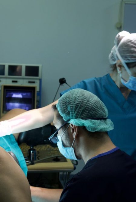
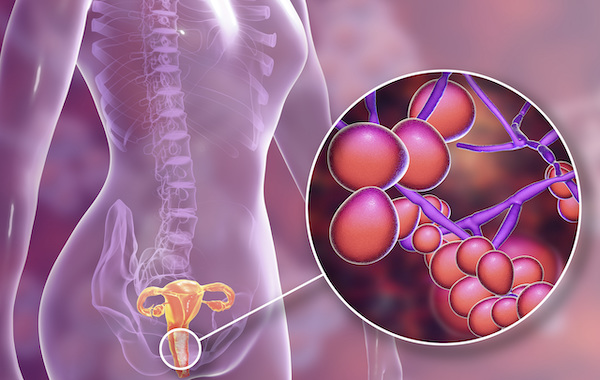
Hysteroscopy procedure is conducted in order for gynecologists to get a clearer idea of what is inside of a patient’s uterus, inserted inside via a hysteroscope through the cervix. The hysteroscope is a thin metal rod that has a light and camera attached in one end, and reflects the image on the camera onto a large screen that the doctor looks at to guide them as they move the instrument.
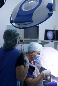 Dr. HIT preparing to insert the hysteroscope inside the cervix. The screen that reflects the instrument’s camera view can be seen in the back.
Who Needs a Hysteroscopy? What are Fibroids in the Uterus and How Do They Affect Fertility?
It is key to understand that not all patients that will receive fertility treatments will require a hysteroscopy. Abnormal bleeding (not during menstruation), pain, and unexplained infertility contribute to the specialist doctor to request for you to receive this operation. Patients who have experienced more than one miscarriage qualify as well, to view if possible blockages like scar tissue on the uterine walls are the reason.
A fibroid is the growth of muscle and tissue that is non-cancerous. If they are not asymptomatic, and cause a considerable amount of discomfort, they are removed. For fertility reasons, fibroids may block the fallopian tubes and inhibit the implantation process, and an embryo cannot form and grow. These fibroids may be removed using another metal instrument that is inserted through the cervical canal, making it a Hysteroscopic Myomectomy.
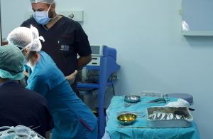
An image showing the medical tools that are kept nearby during the procedure.
How is a Hysteroscopy Done? What is Required Before the Operation? How Does a Hysteroscopy Feel?
This operation is usually not longer than forty-five minutes, and much shorter if it is only a diagnostic one, where no removal of tissue or anything takes place. It is done after the patient undergoes a series of assessments, such as a blood test and pregnancy test, to make sure that they are not pregnant. It is also important to not schedule the hysteroscopy during the days that they are menstruating. The level of pain the individual experiences varies from person to person, although a sedative is given to reduce the discomfort. The area of your vagina and cervix is first disinfected. The doctor opens up the cervix using a bivalve speculum tool, and the hysteroscope rod enters inside. Any abnormalities within the uterus are identified and considered if removal is necessary. The patient is able to leave the hospital the same day after the operation.
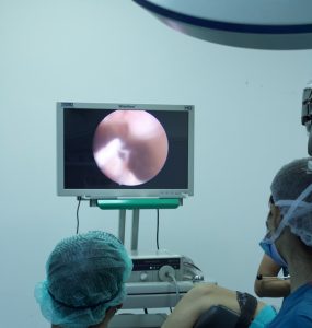 Dr. Erman being guided by the monitor as the hysteroscope moves around the uterus to identify any abnormalities.

